Akshaya Ganji, Adlai E. Stevenson High School (UnknownNow)
Source:1
Introduction
Initially, scientists observe the activation of brain cells in efforts to study the activity patterns in brains. However, more discussion regarding to how long and when these neurons will turn off/on is increasing exponentially2.
Currently, scientists at Scripps Research developed a new technology that allows for a process called inhibition, where it lets them track when, after a burst of activity, brain cells shut off3. Inhibition is the body’s way of regulating gene expression and activity through the usage of operons and other transcription factors, ligands, etc4.
Studies
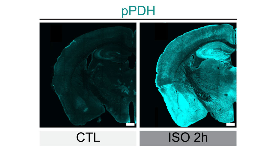
This allows studies to be done regarding how the brains “off switches” go astray, leading to neurological disorders. Through the usage of optogenetics and identifying changes in the pyruvate dehydrogenase (PDH) protein, scientists can now observe how the brain conserves energy by rapidly shutting off this protein post-neuron firing. Optogenetics, a technique in where specific cells are bioengineered to include light-sensitive proteins that act as switches, allows for researchers to precisely control the electrical activity of neurons and nerve cells5.
Moreover, researchers from Duke-NUS Medical School have newly founded a set of light-sensitive proteins that have the capability to turn brain cells off, with just light6.
This team illustrated how through the usage of kalium channelrhodopsins, a type of potassium channel, serves as an effective instrument in regulation of brain gene expression in three experimental animals: fish, worms, and flies.
Potassium ions are essential for electrical function as they maintain cellular processes, such as critical to nerve-impulse transmission, muscle contraction, and cellular-fluid balance maintenance. Dr Stanislav Ott, Senior Research Fellow with Duke-NUS’ Neuroscience and Behavioural Disorders Programme and first author of the study, said: “These potassium channels act like tiny gates on cell membranes. When exposed to light, these gates open and let potassium ions flow through, helping to quiet the activity in the brain cells. This offers us new insights into how brain activities are regulated.”
When triggered by light, the new kalium channelrhodopsins change the electrical gradient across the membrane. This act of hyperpolarization makes it difficult for the neuron to generate action potentials, leading a neuron communication to be suppressed or silenced7.
Future Implications
The action of inhibition through the usage of light-triggered potassium channels studies of interactions between brain regulations, and pathological mechanisms underlying neurodegenerative, neurodevelopmental, and psychiatric disorders.
References
1. New technology lets researchers track brain cells’ ‘off switches’. https://www.scripps.edu/news-and-events/press-room/2024/20240123-le-neurons.html.
2. Singer, E. A Light Switch for the Brain. MIT Technology Review https://www.technologyreview.com/2007/04/05/226043/a-light-switch-for-the-brain/ (2007).
4. Neuroscience News. Illuminating Brain Inhibition. Neuroscience News https://neurosciencenews.com/neural-inhibition-neurotech-25518/ (2024).
6. Lght-controlled ‘off switch’ for brain cells. ScienceDaily https://www.sciencedaily.com/releases/2024/05/240524115302.htm.

Leave a comment